Figure 3.

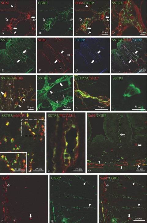
Immunocytochemical detection of SSTRs and several peptides in the non-inflamed and 8w p.i. SSTR4–/– ileum. A–C: Whole-mount preparation of the non-inflamed SSTR4–/– myenteric plexus showing the expression of SOM in the soma of myenteric neurons (filled arrows) and in a vast network of SOM-ir nerve fibres, some of which co-express CGRP (hollow arrows). The arrowheads point towards SOM+/CGRP− nerve fibres. D: SSTR1 was detected in nerve fibres in myenteric whole-mount preparations of the non-inflamed SSTR4–/– ileum. E–H: Immunocytochemical triple staining for SSTR1, SOM and CGRP on cryosections of the non-inflamed SSTR4–/– ileum. All SSTR1-expressing nerve fibres in the lamina propria (filled arrows, E) also expressed SOM (F) and CGRP (G). The hollow arrows in F depict SOM-ir nerve fibres which do not express SSTR1 or CGRP. I–K: Immunofluorescent detection of SSTR2A in cryosections (I) and whole-mount preparations (J, K) of 8w p.i. SSTR4–/– ileum. I: Cryosections revealed SSTR2A expression in S100-labelled glial cells in the myenteric plexus (filled arrow), in the external muscle layers (hollow arrows), in the submucous plexus (filled arrowhead) and in the lamina propria (hollow arrowhead). J: SSTR2A expression in both myenteric neurons (filled arrows) and myenteric glial cells (hollow arrows). K: In the submucous plexus, SSTR2A was only detected in glial cells. L: SSTR3 immunoreactivity (IR) in epithelial cells at the top of the villi as detected in cryosections of non-inflamed SSTR4–/– ileum. M: In the 8w p.i. SSTR4–/– intestine, SSTR3 was observed in almost all mMCP1-labelled mucosal mast cells. The inset shows a higher magnification picture of the white dotted box. N: Endothelial cells of a small-diameter blood vessel in the submucosa, labelled by PECAM-1, showing SSTR5 IR. O: Cryosection of the non-inflamed SSTR4–/– ileum showing widespread distribution of substance P (SubP) and CGRP. Substance P was detected in nerve fibres at the base of the crypts and in the external muscle layers (filled and open arrowheads) and in endocrine cells (filled arrow). The nerve fibres indicated by the hollow arrow and the filled arrowheads did not co-express CGRP, whereas some nerve fibres in external muscle layers contained both substance P and CGRP (hollow arrowhead). The double arrow points to a CGRP+/substance P− nerve fibre in the lamina propria. P-R: Cryosection of the 8w p.i. SSTR4–/– ileum demonstrating substance P IR in endocrine cells (filled arrows) and in nerve fibres in the outer muscle layers (hollow arrow). The filled arrowheads show sprouting of CGRP-ir nerve fibres in the lamina propria. The parasite egg and the surrounding granuloma are indicated by asterisks.
