
Figure 4.
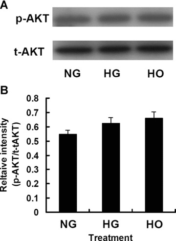
The expression and activation of Akt in rMAPCs were not altered by high glucose or hyperosmolarity after 48 hrs incubation as analysed by Western blot. (A) Representative Western blot for Akt (both total and phosphorylated). (B) Bar graph showing the relative band intensity of the tyrosine-phosphorylated Akt. Results represented the mean ± S.E. of 4 independent experiments. NG: rMAPCs were incubated in the media with 5.5 mM D-glucose; HG: rMAPCs were incubated in the media with 30 mM D-glucose; and HO: rMAPCs were incubated in the media with 24.5 mM mannitol.