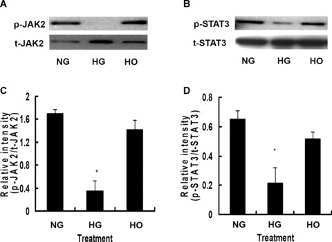
Figure 5.

High glucose significantly inhibited the phosphorylation of JAK2 and STAT3 in rMAPCs after 24 hrs of incubation. (A) Tyrosine phosphorylation of JAK2 was dramatically suppressed by high glucose independent of hyperosmolarity as evaluated by Western blot analysis. The bar graph (C) showed the relative band intensity of phosphorylated JAK2. (B) Phosphorylation of STAT3 was markedly blocked by high glucose independent of hyperosmolarity as evaluated by Western blot analysis. The bar graph (D) showed the relative band intensity of phosphorylated STAT3. NG: rMAPCs were incubated in the media with 5.5 mM D-glucose; HG: rMAPCs were incubated in the media with 30 mM D-glucose; and HO: rMAPCs were incubated in the media with 24.5 mM mannitol. *P < 0.01 compared with NG (5.5 mM D-glucose), n= 4.
