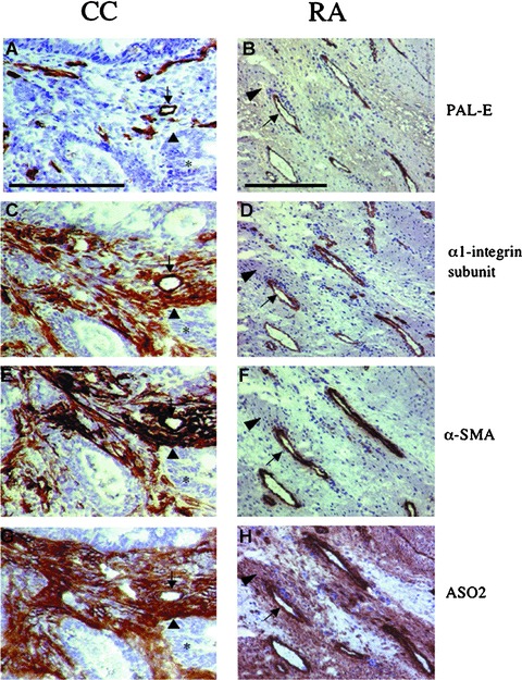
Figure 2.

Expression of phenotypical markers in colorectal adenocarcinoma (CC) and pannus formation in rheumatoid arthritis (RA). Immunohistochemical staining was performed in 6-μm sections from CC (left column) and RA (right column) using mAbs to characterize expression profiles of phenotypical markers in relation to the vasculature for PAL-E (A, B), α1β1 (C, D), α-SMA (E, F) and ASO2 (G and H). Expression profiles were similar in microvascular structures (arrow) in both CC and RA. However, expression profiles differed in interstitial structures (arrowhead) between the two conditions. In CC, the expression profiles in interstitial structures were positive for α1β1 (C), α-SMA (E) and ASO2 (G). In contrast, the expression profiles in interstitial structures in RA were negative for α1β1 (D) and α-SMA (F), but positive for ASO2 (H). Tumour acinar structures (*). The bar represents 200 μm.
