Figure 5.

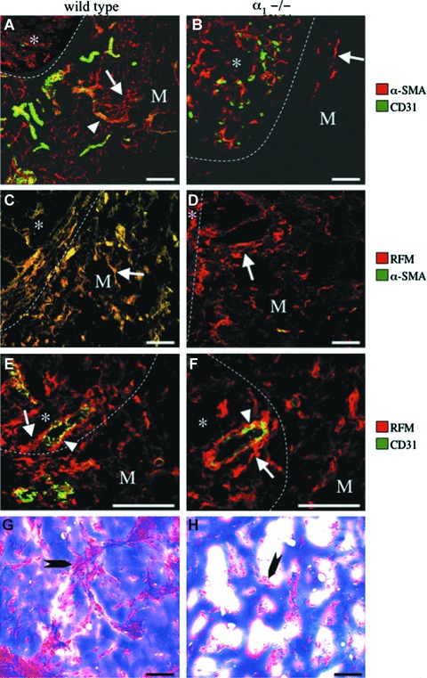
Deficient myofibroblast development and neoformation of blood vessels and supporting connective tissues in the in vivo Matrigel plug assay in α1−/− mice. Double immunofluorescence (A–F) staining using combinations of the mAbs, α-SMA, RFM and CD31, and trichrome (G–H) staining were performed on sections of Matrigel plugs together with adjacent skin that had been grown in the subcutaneous space in wild-type (WT; left column) and α1−/− (KO; right column) mice. Optical sections were obtained using confocal laser microscopy and viewed as composites (A–F). Co-localization is depicted in yellow. (A, B) Note ingrowth into the Matrigel (M) of CD31-positive microvessels (arrowhead) surrounded by α-SMA-positive myofibroblasts (arrow) in wild-type (A) but not in α1−/− mice (B). (C, D) Note ingrowth into the Matrigel (M) of myofibroblasts positive for both RFM and α-SMA in wild-type (C) mice in contrast to Matrigels in α1−/− (D) mice that contained RFM-positive fibroblasts that were negative for α-SMA (arrow). (E, F) Note RFM-expressing cells that are juxtapositioned to, partially detached (arrow) from, the CD31-positive endothelium (arrowhead) that have accumulated in the perivascular space in dermis (*) adjacent to the Matrigel (M) in wild-type (E) mice and, albeit to a lesser degree, also in α1−/− (F) mice. (G, H) Note well-developed cellular connective tissue septa (arrow) in Matrigels in wild-type (G) but not in α1−/− (H) mice, where only isolated accumulations of cells (arrow) were observed. Matrigel/dermal interface (dotted line). The bar represents 40 μm.
