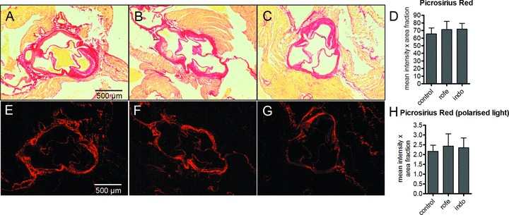
Figure 4.

Collagen content of aortic root lesions of ApoE-deficient mice indicated by picrosirius red staining after treatment with COX inhibitors. Total collagen was evaluated by light microscopy (A–D) and collagen arrangement was analyzed by birefringence analysis using polarized light (E–H). A, E, controls; B, F indomethacin (8 weeks, 3 mg/kg/day); C, G, rofecoxib (8 weeks, 50 mg/kg/day); densitometric quantitation of D, picrosirius red staining, light microscopy and H, picrosirius red staining under polarized light to visualize densely packed collagen (bright red); n= 12, mean ± S.E.M., *P < 0.05.
