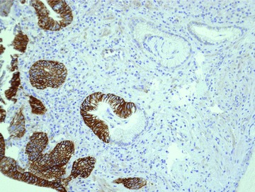
Figure 2.

Area of metaplasia (Barrett’s oesophagus) and dysplasia stained for HER-2 by immunohistochemistry. It is possible to recognize the different expression of HER-2: completely negative in BO and overexpressed in the areas of dysplasia. Original magnification, ×40).
