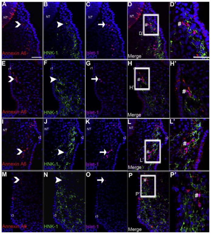
Figure 3. Annexin A6 is absent in migratory neural crest cells but present in the placode cells contributing to the developing trigeminal and epibranchial ganglia at HH13.
Representative transverse sections taken through the chick trigeminal (A–D′), geniculate (E–H′), petrosal (I–L′), and nodose (M–P′) ganglia at HH13 followed by immunohistochemistry for Annexin A6 (A, E, I, M, red), HNK-1 (B, F, J, N, green) and Islet-1 (C, G, K, O, purple), with corresponding merge images shown (D, H, L, P). Carets, arrowheads, and arrows indicate Annexin A6-, HNK-1-, and Islet-1-positive cells, respectively. Annexin A6- and Islet-1-double-positive cells are shown by # in the merge images (D, H, L, P; higher magnification shown in (D′, H′, L′, P′). HNK-1-positive migratory neural crest cells (arrowhead) in the trigeminal (B), geniculate (F), petrosal (J), and nodose (N) ganglia are all Annexin A6-negative. DAPI (blue) labels cell nuclei. Scale bar, 50μm (A–L) and 125μm (C′–L′). Rhombomeres 1 and 2 are denoted by r1 and r2, respectively.

