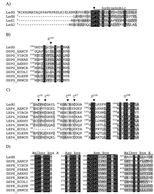
FIG. 3.
Partial protein sequence alignments. (A) Alignment of G. diazotrophicus pseudopilin N terminus. The arrowhead indicates the putative beginning of mature pilins. (B, C, and D) Alignment of LsdG, LsdO, and LsdE with their homologues in other type II secretion systems. S147 in LsdG, C39, C42,C 64, C67, D124, and D188 in LsdO, and Walker A, Walker B, and aspartic acid boxes in LsdE are indicated above the alignments. The sequences, identified by their SwissProt entry names, correspond to the following species: Xanthomonas campestris, Vibrio cholerae, P. aeruginosa, Aeromonas hydrophila, Erwinia carotovora, E. coli, Klebsiella pneumoniae, and Erwinia chrysanthemi. Identical and similar residues are indicated by white letters on a black background and by black letters on a gray background, respectively. Groups of residues considered similar in the alignment are described in Table 3, footnote a. The numbers in the alignment indicate the positions of the residues in the precursor proteins.

