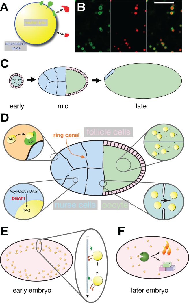
Figure 1. The life cycle of Drosophila embryonic lipid droplets.
(A) Structure of lipid droplets: A core of neutral lipids (triglycerides (TAG), sterol esters, retinol esters) is surrounded by a monolayer of amphipathic lipids (such as phosphoglycerides and sterols). Proteins can be stably embedded (green) in this monolayer or reversibly bound (red) to other proteins or lipid head groups. (B) Colocalization of a GFP fusion (green) that targets to lipid droplets (GFP-LD [56]) and neutral lipids (red, detected with the dye Nile Red). The fusion protein surrounds a core of neutral lipids. Note that this particular fusion protein labels only a subset of lipid droplets. Scale bar = 5 μm. Image modified from [56]. (C) Overview of oogenesis (for color code of cell types, see D). In early egg chambers, the 16-daughter cells of the germ-line cytoblast (the future nurse cells and oocytes) are surrounded by a layer of somatic follicle cells. In mid-stage egg chambers, nurse cells supply the growing oocyte with nutrients, proteins and RNAs; follicle cells have migrated to cover the oocyte. In late stages, nurse cells have transferred most of their contents to the oocyte and have undergone apoptosis. The oocyte is surrounded by an eggshell (not shown) that was produced by the follicle cells. The various stages are not drawn to scale; by the end of oogenesis the oocyte volume has increased more than a hundred fold. (D) Lipid droplets in mid-stage egg chambers. One of the ring canals connecting nurse cells and oocyte is indicated. Top left: lipophorin particles (diacylglycerol (DAG) rich components of the hemolymph) are taken up by nurse cells via lipophorin receptors (Lpr). Bottom left: in the nurse cells, DGAT1 converts acyl-CoA and DAG into TAG, which contributes to the growth of the LD core. Bottom right: cytoplasmic streaming transports lipid droplets from nurse cells through ring canals into the oocyte. Top right: in the oocyte cytoplasm, most lipid droplets move passively by cytoplasmic streaming; a subset is actively transported by motors along microtubules. (E) In early embryos, lipid droplets move bidirectionally along microtubules. (F) Later in embryogenesis, lipid droplets are thought to be broken down to generate energy (via oxidative phosphorylation) and building blocks (for biomass production).

