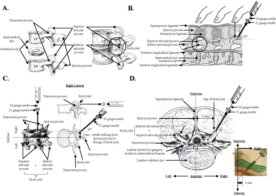
Fig. 1.
Schematic diagram for the induction of facet joint osteoarthritis by percutaneous needle puncture. A, The anatomical structure of a facet joint of the lumbar spine. Representative image is from http://www.elu.sgul.ac.uk/rehash/guest/scorm/193/package/content/lumbar_vertebra_lateral_view.htm. B, Facet joint localization in the rat by palpation of the back. Representative image is from Joseph E. Donnelly, 1982, Living Anatomy, (Champaign, IL: Human Kinetics), 87. C, Representation of percutaneous needle puncture of facet joint capsular tissues using a combination of 21-gauge and 26-gauge needles. Images are from http://www.columbianeurosurgery.org/2010/02/back-pain-anatomy-lesson/ and http://morphopedics.wikidot.com/fractures-of-the-l4-l5-vertebrae. D. Horizontal view of the percutaneous needle puncture of facet joint capsular tissues performed to induce facet joint osteoarthritis. Representative image is from Brown JE, Nordby EJ and Smith L, Chemonucleolysis, Thorofare, NJ, 1985, Slack.

