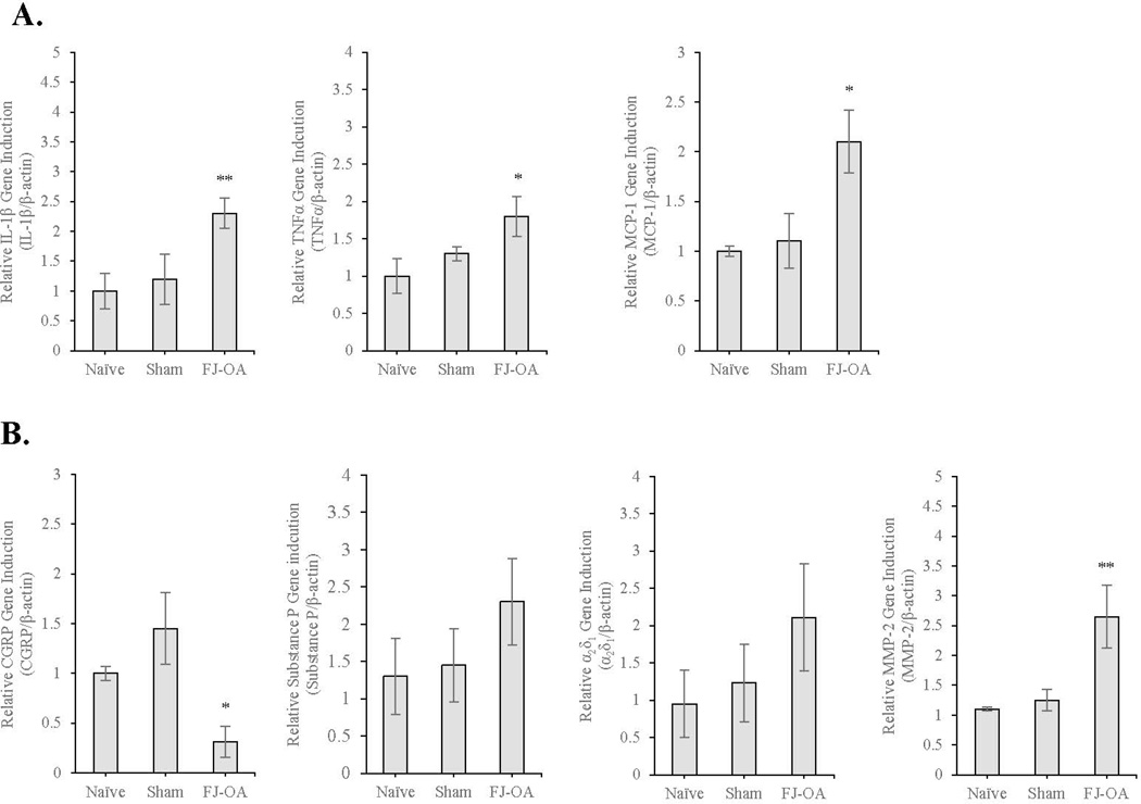
Fig. 8.
Gene expression analyses by qPCR using the spinal cord dorsal horn of the percutaneous needle puncture-induced FJ OA. A. Compared to sham-operated (n=3) and naïve controls (n=3), there was a significant increase in IL-1β (**p<0.01), TNFα (*p<0.05), and MCP-1 (*p<0.05) in the FJ OA model. B. There was a decrease in CGRP (*p<0.05) and an increase in MMP-2 (**p<0.01) in the FJ OA model. Expression of substance P (p=0.50832) and α2δ1 (p=0.42285) was slightly increased without statistical significance. No difference was observed among naïve and sham-operated controls. Each value represents the mean ± SE. IL-1β=interleukin-1β; TNFα=tumor necrosis factor α; MCP-1=monocyte chemoattractant protein-1; CGRP=calcitonin gene-related peptide; MMP-2=matrix metalloproteinase-2

