
Figure 1.
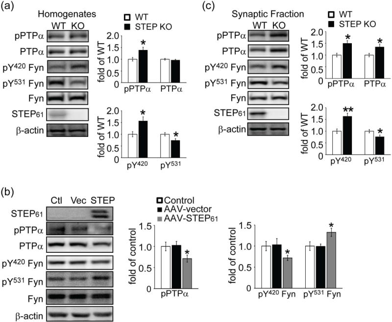
Phosphorylation level of PTPα at Tyr789 is elevated in STEP KO mouse striatum and decreased when STEP61 is restored to STEP KO cultures. Tyrosine phosphorylation levels of PTPα (pY789), Fyn (pY420 and pY531), and total protein levels were determined in total homogenates from wild-type (WT) and STEP KO (KO) mouse striatum (a), corticostriatal neurons from STEP KO mice with restoration of STEP61 expression using AAV1/2-STEP61 (b) or synaptic membrane fractions from wild-type (WT) and STEP KO (KO) mouse striatum (c) Quantification of phosphorylation levels were normalized to total protein levels and then to β-actin as a loading control. All data were expressed as mean ± SEM (*p < 0.05, **p < 0.01, Student’s t-test for (a) and (c), one-way ANOVA with Tukey’s test for (b); n = 6).