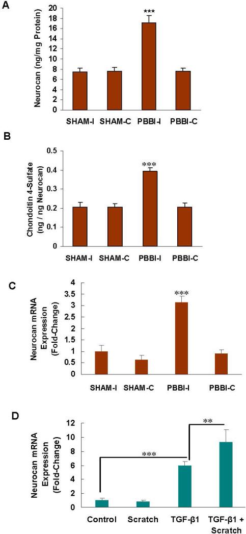
Fig. 4. Increase in neurocan and in C4S immunoprecipitated neurocan following injury.
A. Neurocan, as measured by ELISA, showed significant increase in the ipsilateral traumatized brain tissue (p<0.001).
B. The C4S that immunoprecipitated with neurocan, expressed as ng C4S per ng neurocan, increased following injury, compared to the SHAM controls (p<0.001) and to PBBI-C (p<0.01). This increase is substantial, since the neurocan content was also increased post-trauma. The increase in C4S is attributable to both impaired degradation due to decline in ARSB and increased production due to higher levels of CHST11 expression and sulfotransferase activity.
C. The neurocan mRNA was significantly increased at 24 hours in PBBI-I (p<0.001).
D. In the astrocytes, the neurocan mRNA increased following TGF-β1 (p<0.001), but not scratch. The combination of scratch and TGF-β1 further increased the neurocan expression over TGF-β1 alone (p<0.01).

