
Figure 6.
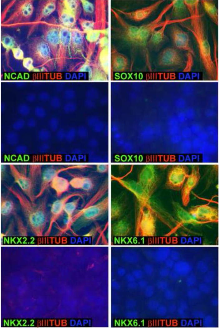
Immunocytochemical confirmation of expression of some DMSO-responsive genes. Marmoset iPS clone B8 cells were incubated for 24 hours in 1% DMSO and then subjected to the 6 day differentiation protocol described in Fig. 1. Cells were plated on glass coverslips for immunocytochemistry with antibodies against NCAD, Sox10, Nkx2.2 and Nkx6.1 (green fluorescence; see Materials and Methods). In each case the cells were also stained with an antibody against βIII tubulin (red) and were costained with DAPI (blue). For each of 4 the genes the upper images are of the cells following differentiation and the lower images are of the undifferentiated iPS cells.