Fig. 1.


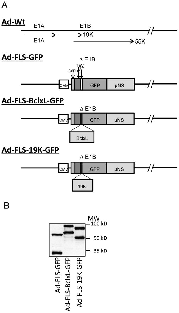
A. Schematic diagram of the E1 region of recombinant hAdv5. The recombinant viruses express the indicated chimeric proteins in the E1B region under the control of CMV-IE promoter. GFP, BCLxL-GFP and E1B19K-GFP are tagged at the N-terminus with Flag, TEV and ST tags and at the C-terminus with the orthoreovirus μNS domain.
B. Expression of chimeric proteins by recombinant viruses. The proteins expressed in infected A549 cells were analyzed by western blotting using Flag antibody and visualized with western blotting detection system.
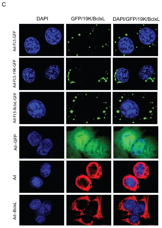
C. Localization of GFP fusion proteins into cytoplasmic inclusion bodies. The infected cells were stained with BCL-xL and E1B-19K antibodies. The immunofluorescence and GFP fluorescence were viewed in several fields and a single representative field in each category was photographed using a confocal microscope.
