
Fig. 3.
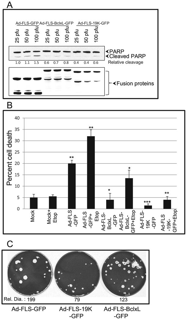
Suppression of apoptosis by chimeric BCL-xL and E1B-19K proteins localized at non-canonical sites. A. A549 cells were infected with the recombinant viruses at different MOI and 24 hr post infection cells were lysed and analyzed by western blotting to determine the extent of PARP cleavage (top panel). The western blot with the Flag Ab (lower panel) shows the levels of expression of different recombinant proteins. B. Effects on apoptosis. A549 cells were infected with different viruses (50 pfu/cell) and cell viability was determined by trypan blue using a BIORAD cell counter. The experiment was repeated three times and the statistical differences were assessed using two-tail student t-test with 95% confident. P-value was determined by comparing mock vs Ad-FLS-GFP, mock+etop vs Ad-FLS-GFP+etop, Ad-FLS-GFP vs Ad-FLS-BCLxL-GFP and Ad-FLS-E1B19K-GFP and Ad-FLS-GFP+etop vs Ad-FLS-BCLxL-GFP+etop and Ad-FLS-E1B19K-GFP+etop. Significance was accepted at a value of *p<0.05, **p<0.01 and ***p<0.005. C. Plaque morphology of recombinant viruses. A549 cells were infected with the recombinant viruses and assayed for plaque formation (Subramanian et al., 2007). The diameters of 24 to 50 plaques were measured using ImageJ and the relative diameter (Rel.Dia.) is shown at the bottom of each dish.