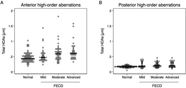
Figure 1.

High-order aberrations (HOAs) at the anterior and posterior cornea in Fuchs endothelial corneal dystrophy (FECD, n = 107) and normal (n = 71) measured with Scheimpflug imaging. Posterior corneal HOAs were of much smaller magnitude than anterior corneal HOAs. (A) Anterior HOAs in moderate (p =0.01) and advanced (p =0.01) FECD were increased compared to controls. (B) Posterior HOAs were increased even in mild (p =0.02), moderate (p <0.001), and advanced (p <0.001) FECD compared to controls. Significance was determined with GEE models adjusted for age. Each dot represents one observation; median is indicated with black line.
