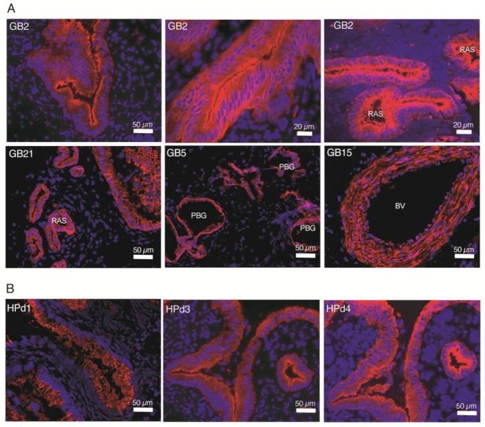
Figure 1. Immunofluorescence labeling patterns in adult human gallbladder.
A) The mAb GB2 labeled mucosal epithelia (top left and top middle panels) with characteristic outpouching (also known as Rokitansky-Aschoff sinuses (RAS)) (top right) extending into the muscularis layer. Also, GB21 selectively labeled mucosal epithelium (bottom left), but without the apical bias of GB2. GB5 mAb marked peribiliary glands (PBGs) (bottom middle). GB15 labeling of a blood vessel (BV) wall (bottom right). B) Three pancreatic ductal surface antibodies labeled gallbladder mucosal epithelium.

