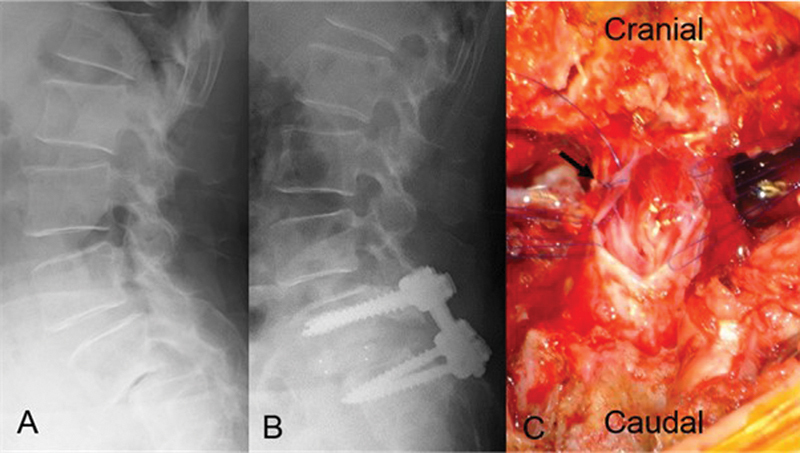
Fig. 1.

Plain lateral radiographs (A, B) and intraoperative photograph (C). (A) Preoperative plain radiograph shows L5 degenerative spondylolisthesis with 47% vertebral slip. (B) Postoperative plain radiograph shows L5 vertebral slippage of 23%. (C) Intraoperative imaging after enlargement of the dural tear. The location of the new dural tear is different from that of the primary dural tear (arrow).
