
Fig. 2.
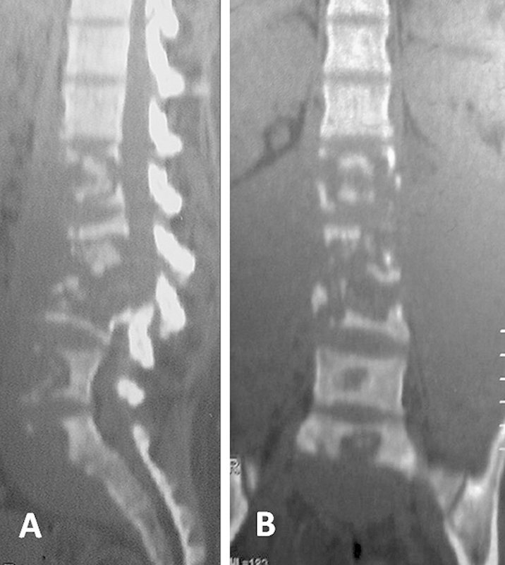
Sagittal (A) and coronal (B) computed tomographic scans showing extensive vertebral body destruction from L1 to upper sacral vertebrae.
Official websites use .gov
A
.gov website belongs to an official
government organization in the United States.
Secure .gov websites use HTTPS
A lock (
) or https:// means you've safely
connected to the .gov website. Share sensitive
information only on official, secure websites.
Sagittal (A) and coronal (B) computed tomographic scans showing extensive vertebral body destruction from L1 to upper sacral vertebrae.