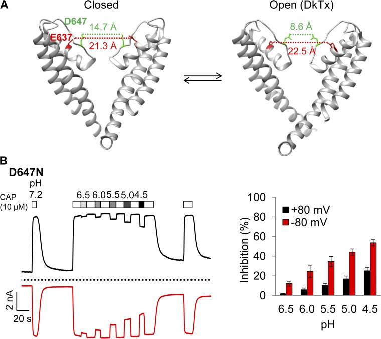
Figure 12.
Concentration- and voltage-dependent inhibition of D647N current by H+. (A) The cryo-EM pore structures of TRPV1 in the closed (left; Protein Data Bank accession no. 3J5P) and double-knot toxin DkTx–bound (right; Protein Data Bank accession no. 3J5Q) states. Only two of the four subunits are shown. Dotted lines indicate the distances between the hydroxyl groups of E637 and D647. (B) Representative current traces induced by 10 µM capsaicin from whole-cell recording at ±80 mV in Solution IV containing different concentrations of H+ (left) and the percentage of current inhibition (right). Dotted line indicates zero current level. n = 4. Error bars indicate mean ± SEM.

