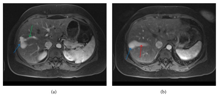
Figure 3.

A 45-year-old female with intrahepatic portosystemic venous shunt with associated aneurysms. (a) Contrast enhanced axial VIBE MRI image showing communication between the right portal vein (green arrow) and right hepatic vein through an aneurysm (blue arrow). (b) Contrast enhanced axial VIBE MRI (a few sections cranial to Figure 2(a)) showing communication between the right portal vein and right hepatic vein (red arrow) through a second larger aneurysm (blue arrow). Protocol: Siemens, 1.5 Tesla Avanto MR Scanner, TR = 4.3, TE = 1.91, 3.5 mm slice thickness, Matrix = 192 × 256, with 15 mL Gadobenate dimeglumine (Multihance, Bracco Diagnostics Inc.) injection.
