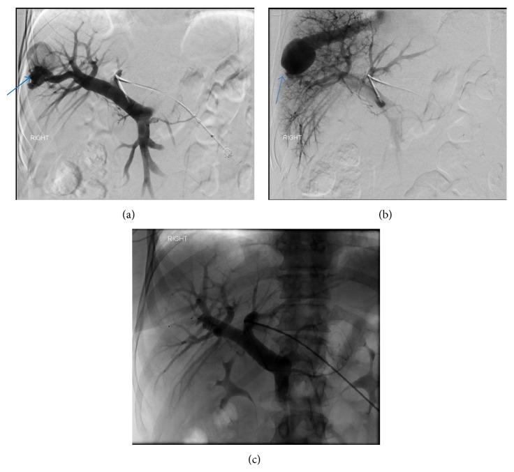
Figure 5.
A 45-year-old female with intrahepatic portosystemic venous shunt with associated aneurysms. (a) Portal venography obtained via left portal vein with catheter tip in the main portal vein demonstrates a right anterior portal vein branch communicating with the right hepatic vein through a 28 mm aneurysm (arrow). (b) Portal venography obtained via left portal vein with catheter tip in the main portal vein shows a right posterior portal vein branch communicating with the right hepatic vein through a second, larger, 37 mm aneurysm (arrow). (c) Portal venography performed after embolization of both anterior and posterior right portal branches communicating with the right hepatic veins with Amplatzer vascular plugs demonstrates obliteration of intrahepatic portosystemic shunts. Protocol: Single Plane Artis Zee Siemens system, embolization with a 12 mm and 6 mm Amplatzer vascular plug (St Jude Medical).

