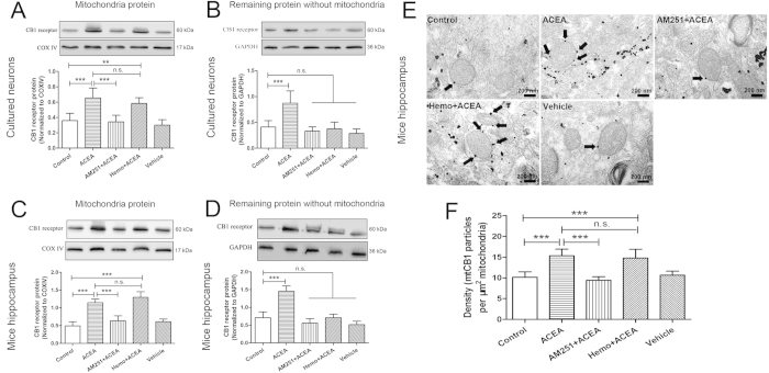
Figure 2. Expression of CB1 receptor protein in primary cultured hippocampal neurons and mouse hippocampus.
(A, C) Western blot showing mtCB1R protein expression at 2 h after administration in vitro and in vivo (n = 5). (B, D) Western blot showing CB1 protein expression in the remaining samples without mitochondria protein at 2 h after administration in vitro and in vivo (n = 5). (E) Immunogold electron microscopy showing mtCB1R protein expression. (F) Density of mtCB1R immunoparticles were calculated per area of mitochondria (n = 3). Scale bars = 200 nm. Data represent mean ± SD. **P < 0.01; ***P < 0.001; n.s.: no significance. COX IV: cytochrome c oxidase; Hemo: hemopressin.

