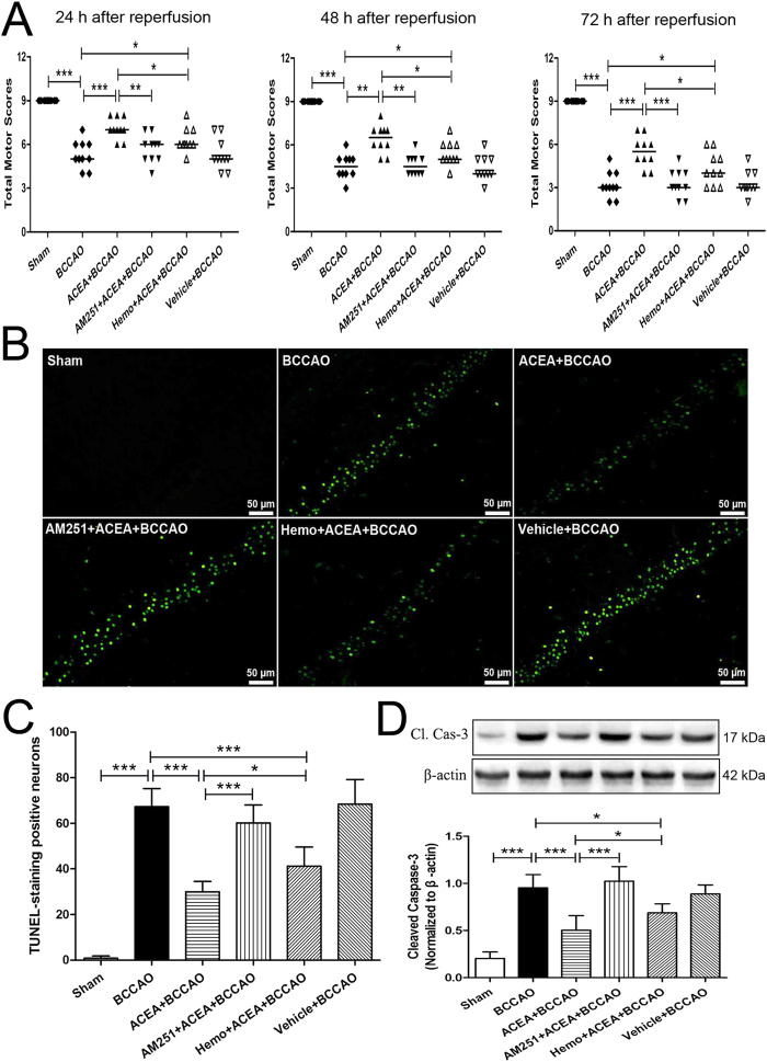
Figure 5. ACEA-induced neuroprotection against cerebral ischemia/reperfusion injury in mice.
(A) Neurological assessment at 24 h, 48 h and 72 h after reperfusion (n = 13). Data represent the median. (B) Representative photomicrographs of TUNEL staining in hippocampal CA1 region. Scale bars = 50 μm. (C) Quantitative analysis of the number of TUNEL-positive cells in hippocampal CA1 region at 72 h after reperfusion (n = 5). (D) Western blot showing representative the expression of cleaved caspase-3 (Cl. Cas-3) protein in the hippocampus at 72 h after reperfusion (n = 5). Data represent mean ± SD. *P < 0.05; **P < 0.01; ***P < 0.001. BCCAO: bilateral common carotid artery occlusion; Hemo: hemopressin.

