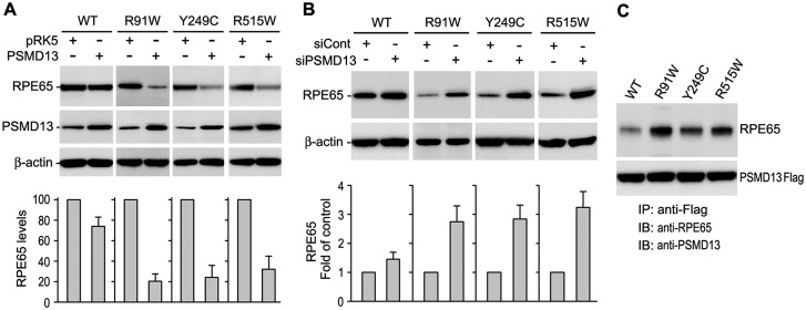
Fig. 2.
PSMD13 mediated degradation of mutant RPE65 proteins. (A) Immunoblot analysis of WT or the indicated mutant RPE65 in ARPE-19 cells co-transfected with pRK5 mock vector or PSMD13 construct. PSMD13 expression was monitored by immunoblot analysis; β-actin was used as a loading control. Relative intensities of the RPE65 immunoblots were quantified and expressed as percentage of WT RPE65 in the histograms. (B) Immunoblot analysis showing increase in expression levels of the mutant RPE65s in ARPE-19 cells co-transfected with PSMD13 siRNA (siPSMD13). Histogram shows relative expression levels of RPE65 in the cells. (C) Immunoprecipitation showing strong interaction of PSMD13 with mutant RPE65 proteins. The 293 T-LC cells expressing PSMD13-Flag fusion protein and WT or mutant RPE65 were immunoprecipitated with a Flag antibody and the precipitates were probed with antibodies against RPE65 or PSMD13.

