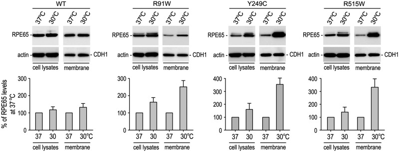
Fig. 7.
Increased membrane-association of the mutant RPE65s at low temperature. Total cell lysates and membrane fractions of 293 T-LC cells expressing WT or mutant RPE65 were analyzed by immunoblot analysis using antibodies against RPE65, β-actin or E-cadherin (CDH1). Histograms show relative contents of RPE65 proteins in the indicated fractions of cells maintained at 37 versus 30 °C. Contents of RPE65 in the cell lysates and membrane fractions were normalized by β-actin or CDH1 levels, respectively. Error bars show SD (n = 3).

