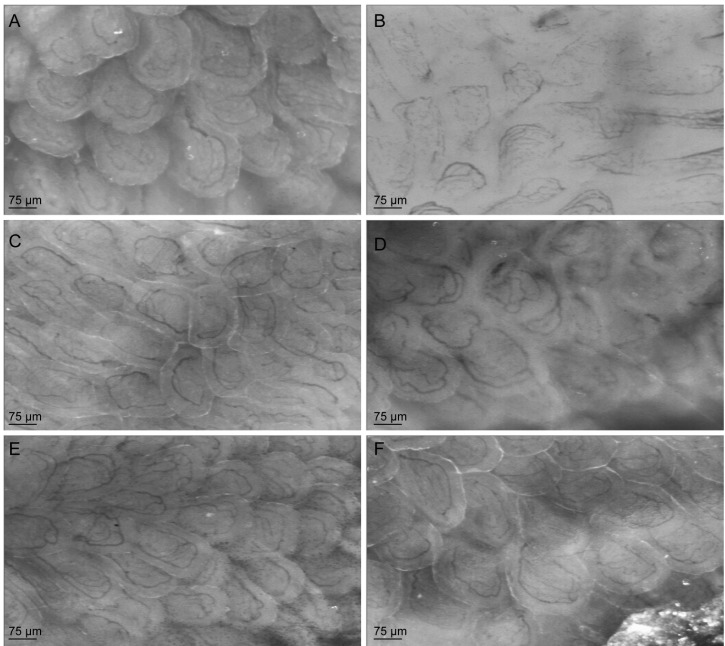
Figure 10.
Representative live images of the microcirculatory measurements in jejunal villi with side stream dark-field (SDF)-imaging of control and arginase treated animals with or without l-citrulline or l-arginine supplementation. (A) Representative image of the jejunal microcirculation in a control mouse. (B) Representative image of an arginase-treated mouse, with a decreased number of perfused vessels per villus. (C) Representative image of the jejunal microcirculation in an l-arginine-treated mouse, which shows a comparable perfusion pattern as the control mouse. (D) Representative image of an arginase + l-arginine-treated mouse, which shows no beneficial effect of l-arginine supplementation on the perfusion. (E) Representative live image of an l-citrulline-treated mouse, which also shows a comparable perfusion pattern as the control and l-arginine-treated mouse. (F) Representative image of an arginase + l-citrulline-treated mouse, which shows more perfused vessels per villus compared to arginase and arginase + l-arginine-treated animals.

