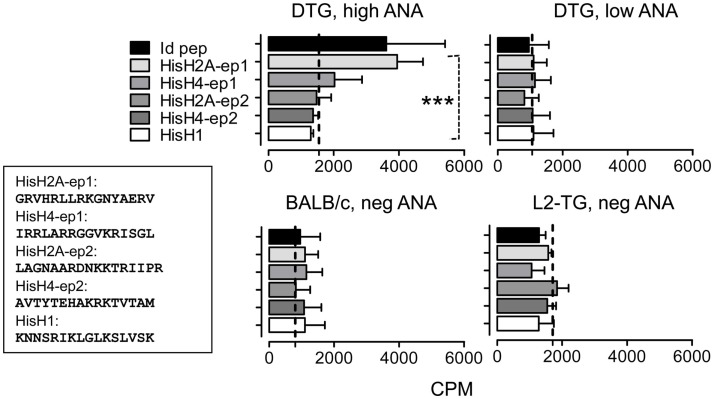
Figure 5.
Analysis of histone-specific Th cell responses in DTG mice and controls. Based on the predictions in Figure 4 and Figure S1 in Supplementary Material, histone peptide stretches with similarities with mouse IgVH CDR3 sequences were tested in DTG lupus mice. Lymph node Th cells were from controls (bottom) or high and low serum ANA DTG mice were tested for responses toward Id-peptide, or histone peptides from HisH1, HisH2A, or HisH4 (see Materials and Methods and Figure 4 where His H2A and H4 peptides are shown) presented by irradiated BALB/c splenocytes. Dotted line: Control without peptide, n = 6. Upper left histograms (DTG, high ANA): One-way Anova, p < 0.0008, with Tukey’s Multiple Comparison test, p < 0.05 for His2A-ep1 vs. other histone peptides and control).

