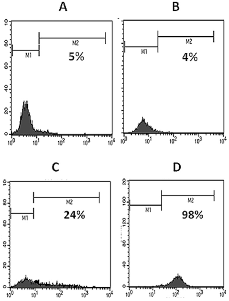
Figure 3 .

Flow cytometric analysis of expression of human CD31. Using specific monoclonal antibody, expression of human CD31 on NIH-3T3 (A), mock-transfected NIH-3T3 (B) and pCMV6-Neo/CD31-transfected NIH-3T3 cells (C, for transient and D, for stable expression) were analyzed.
