
Figure 3 .
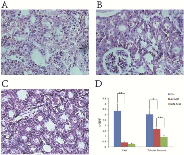
Renal histology following transplantation of Nrf2-MSCs and Ad-MSCs. A, B and C; Light microscopy images of renal tissue from rats (H&E stained kidney sections, 400×). A; Cisplatin group: tubules showed extensive and marked changes consisting of flattening, loss of epithelial cells and brush border. B; Ad-MSC group: less severe tubular damage and very mild cell swelling were observed. C; Nrf2-MSC group: higher recovery and similarity to normal morphology of kidney. D; Renal histology (casts and tubular necrosis, quantified as number per high power field (HPF)) of cisplatin treated rats. Tubular necrosis and lesions included loss of brush border and epithelial cells, flattening, , and luminal cell debris. (Mean±SE, *: P<0.05, **: P<0.01 and ***: P<0.001)