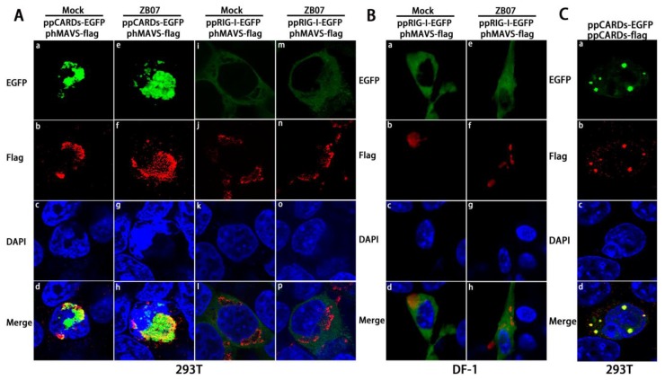
Figure 4.
Co-localization of pigeon CARDs with MAVS (A) phMAVS-flag and ppRIG-I-EGFP/ppCARDs-EGFP were co-transfected into 293T cells respectively, and 24 h later, transfected cells were infected with ZB07 viruses or mock-treated, then, transfected cells were dyed with anti-flag-antibody and DAPI at 8 h p.i. Then, the cellular localization of pRIG-I, CARDs, and hMAVS was examined via confocal microscopy; (B) pcMAVS-flag and ppRIG-I-EGFP were co-transfected into DF-1 cells respectively, and 24 h later, transfected cells were infected with ZB07 viruses or mock-treated, then, transfected cells were dyed with anti-flag antibody and DAPI at 8 h p.i. Then, the cellular localization of pRIG-I and hMAVS was examined via confocal microscopy; (C) 293T cells were co-transfected with ppCARDs-EGFP and ppCARDs-flag. 24 h later, transfected cells were dyed with DAPI, anti-flag antibody, and cy3 labled secondary antibody. Then, the cellular localization of pCARDs-EGFP and CARDs-flag was examined via confocal microscopy.

