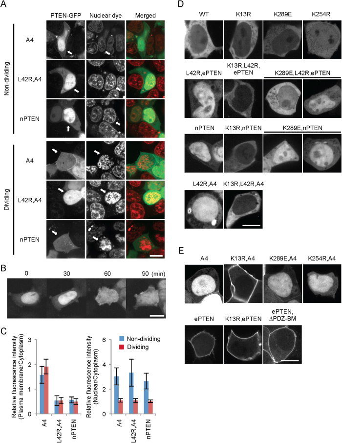
Figure 2. PTENA4 is redistributed to the plasma membrane in the absence of the nuclear membrane during mitosis.
(A) HEK293 cells expressing the indicated versions of PTEN-GFP were stained with a DNA dye, DRAQ5 (Cell Signaling), and observed by fluorescence microscopy. Dividing and non-dividing cells were identified by nuclear staining. Bar, 10 μm. (B) HEK293 cells expressing PTENA4-GFP were observed by time-lapse confocal microscopy. PTENA4-GFP was translocated to the plasma membrane upon nuclear membrane breakdown, which happened between 30 and 60 min. (C) Intensity of GFP at the plasma membrane and in the nucleus was quantified relative to that in the cytosol. Values represent the mean ± SD (n ≥ 8). (D,E) K13R blocks the nuclear accumulation of PTEN with the open conformation. HEK293 cells expressing the indicated forms of PTEN-GFP were observed by fluorescence microscopy. Bar, 10 μm.

