
Fig. 1. Increase in severity of DENV-2 infections in two independent studies of pediatric dengue in Nicaragua.
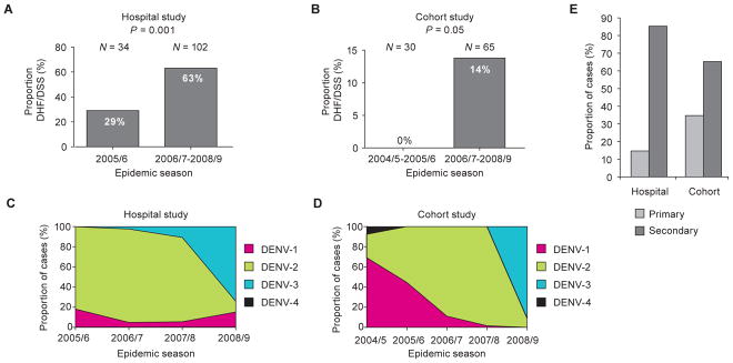
(A, B) The proportion of DENV-2 cases, including both primary and secondary infections, classified as DHF/DSS in the early as compared to later seasons in the Hospital (P = 0.001, Fisher’s exact test) (A) and Cohort (P = 0.05, Fisher’s exact test) (B) studies. The total number (N) of DENV-2 infections included in the analysis is shown above each graph. (C, D) The proportion of each serotype across four epidemic seasons (2005/6–2008/9) in the Nicaraguan Hospital study (C) and five epidemic seasons (2004/5–2008/9) in the Nicaraguan Pediatric Dengue Cohort study (D). Pink = DENV-1; green = DENV-2; blue = DENV-3; black = DENV-4. (E) The proportion of all DENV-2 cases classified as primary (light gray bars) or secondary (dark gray bars) DENV infections in the Hospital and Cohort studies, as defined in Materials and Methods.