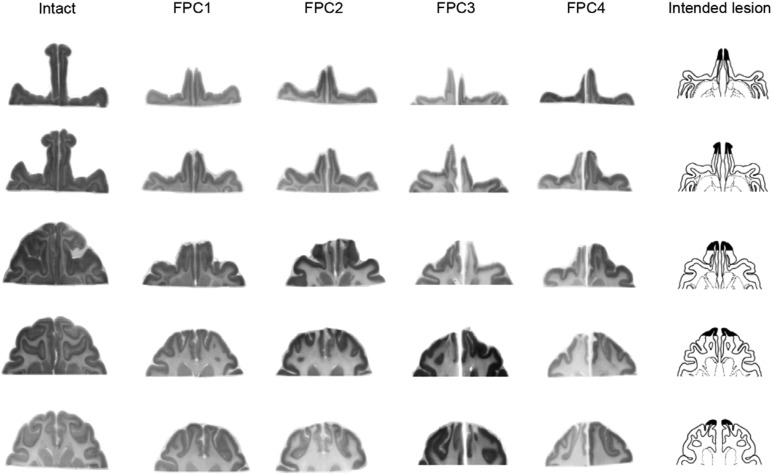
Fig. 2.
Extent of FPC lesions confirmed in horizontal brain sections. Nissl-stained horizontal sections were taken from five dorsoventral levels where FPC had existed in each monkey. The column 1 is for an intact monkey (intact), columns 2–5 are for four FPC-lesioned monkeys (FPC1–FPC4), and column 6 shows the intended lesion extent on drawings of a representative brain. Note the absence of gray matter tissue at the most rostral part of the brain in each FPC-lesioned animal. Microscopic examination of the stained sections confirmed complete lesions of the entire extent of the cortex in the frontal pole, with no damage outside of the intended region.

