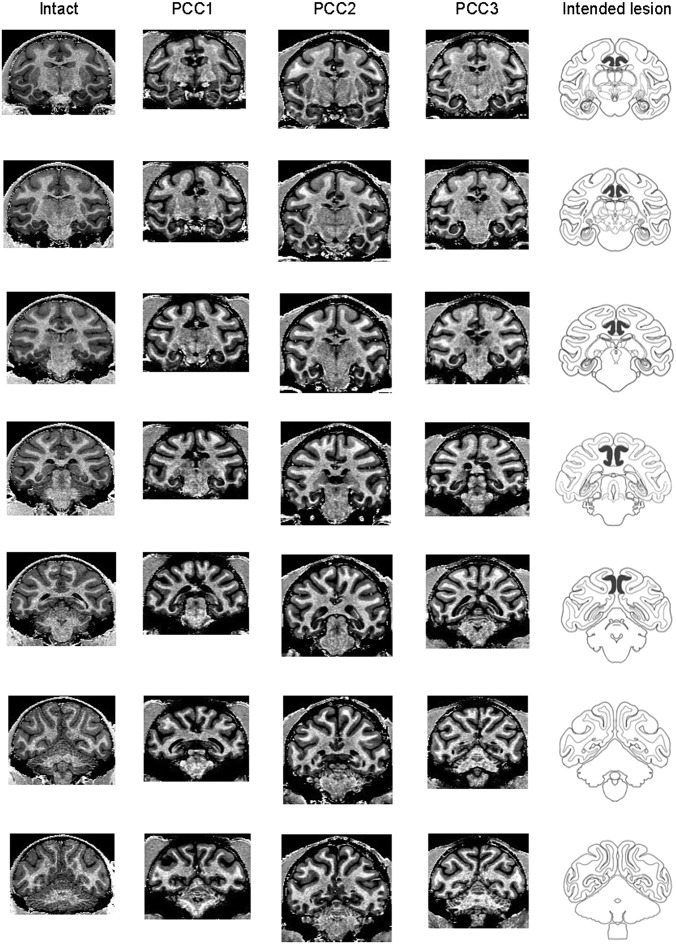
Fig. 3.
Extent of PCC lesions confirmed in MRI. MRI scans were taken after the end of postoperative data collection to verify the lesion extent. Column 1 is for an intact monkey (intact), and columns 2–4 are for three PCC-lesioned monkeys (PCC1–PCC3). Frontal sections are taken from seven anterior–posterior levels covering the PCC in each monkey. The sections in PCC-lesioned monkeys were selected so that their gyri and sulci shapes match those of the intact monkey as much as possible. The gray matter tissues are missing bilaterally in the ventral bank of cingulate sulcus and on the surface of posterior cingulate gyrus in each PCC-lesioned animal. The lesions of PCC were as intended, with no damage outside the target area (Fig. S1). The schematic diagrams showing the intended lesion extent (column 5) were adapted from the NIMH (National Institute of Mental Health) Rhesus Macaque Brain Atlas.

