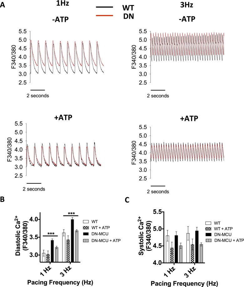
Fig. S7.
Reversal of elevated diastolic cytosolic [Ca2+] in voltage clamp-stimulated DN-MCU ventricular myocytes by ATP dialysis. (A) Representative [Ca2+] transients from isolated ventricular WT (black tracings) and DN-MCU (red tracings) myocytes stimulated at 1 and 3 Hz in the absence or presence of ATP (5 mM), which was added to the pipette (intracellular) solution. Summary data for diastolic (B) and systolic (C) cytosolic [Ca2+] in WT and DN-MCU ventricular myocytes in the presence and absence of pipette solution with 5 mM ATP. Cell number in each group is indicated in parentheses: (B) 1 Hz WT (7), WT + ATP (8), DN-MCU (16), DN-MCU + ATP (11); 3 Hz WT (8), WT + ATP (7), DN-MCU (13), DN-MCU + ATP (9). (C) 1 Hz WT (6), WT + ATP (6), DN-MCU (9), DN-MCU + ATP (6); 3 Hz WT (8), WT + ATP (7), DN-MCU (9), DN-MCU + ATP (10). ***P < 0.001, one-way ANOVA analysis of means. Error bars show SEM.

