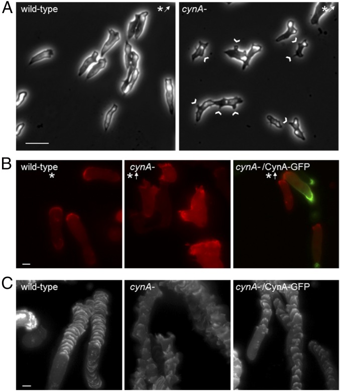
Fig. 5.
Disruption of CynA leads to an increase in lateral protrusions and an altered leading edge morphology. (A) Differentiated wild-type and cynA- cells were imaged by time-lapse microscopy for 30 min at 30-s intervals while migrating toward a micropipette filled with cAMP. Images are shown for the last frame. Asterisks indicate the position of the micropipette; arrows indicate that the micropipette was placed outside of the frame shown. Arrowheads indicate lateral protrusions. (Scale bar, 20 µm.) (B and C) Differentiated wild-type and cynA- cells coexpressing LimEΔcoil-RFP and either CynA-GFP or empty vector were imaged by fluorescence microscopy at 30-s intervals while chemotaxing toward a micropipette filled with cAMP (B). The images shown in B are a merge of the GFP and RFP channels. Tracks of the RFP fluorescence intensities for the cells shown in B were generated using ImageJ, such that each pixel shown in the overlaid image corresponds to the maximum pixel intensity for that location over the course of the movie (C). The first 5 min of each movie, during which cells were turning to orient toward the cAMP gradient, were excluded from analysis in C for simplification. Asterisks indicate the position of the micropipette; arrows indicate that the micropipette was placed outside of the frame shown. (Scale bars, 5 µm.)

