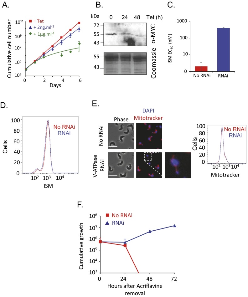
Fig. S3.
A second V-ATPase subunit sensitizes cells to isometamidium. (A) Cumulative growth of V-ATPase RNAi strains for the Vo-d subunit (Tb927.5.550). Error bars indicate SD calculated from two independent strains. (B) Western blot analysis showing depletion of the MYC-tagged d-subunit. The Coomassie panel serves as a loading control. Tetracycline was applied at 2 ng/mL. (C) RNAi-based depletion of the d-subunit renders cells >100-fold resistant to ISM. Error bars indicate SD derived from two independent strains. (D) Flow cytometry analysis of ISM staining following d subunit RNAi. (E) Microscopy and flow cytometry analysis of Mitotracker staining following d-subunit RNAi. (Scale bars, 10 μm.) The magnified image shows the expected thread-like structure. (F) Cumulative growth of RNAi strains after exposure to acriflavine. Error bars indicate SD calculated from two independent strains.

