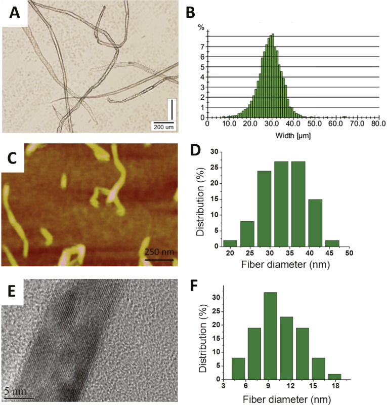
Fig. S2.
(A) Optical microscope image of native cellulose fiber with a mean diameter of 27 μm. (B) Size distribution histogram. (C) AFM image of cellulose fibers with mean diameters of 28 nm. (D) Size distribution histogram. (E) HRTEM crystalline lattice image of fiber with a mean diameter of 11 nm. (F) Size distribution histogram.

