Fig. S3.
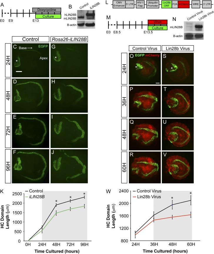
LIN28B overexpression delays auditory HC differentiation. (A–K) Acute overexpression of iLIN28B slows the progression of HC differentiation. (A) Experimental design: Rosa26-iLIN28B overexpression was induced at E12.5, and control and iLIN28B cochlear explants were cultured 12 h later at E13.0. HC differentiation was monitored over the following 4 d by using HC-specific Atoh1/nEGFP reporter expression. (B) Human LIN28B protein is overexpressed within the developing cochlear epithelium (E13.5) of Rosa26-iLIN28B mice 24 h after the start dox treatment. (C–J) Acute overexpression of iLIN28B slows the progression of HC differentiation. Atoh1/nEGFP reporter expression (EGFP, green) marks nascent HCs. Asterisks indicate EGFP expression within HCs of the vestibular sacculus. (K) Length of the EGFP+ HC stripe was used to quantify the extent of HC differentiation in control versus Rosa26-iLIN28B cochlear cultures. The gray box indicates the estimated time-point at which LIN28B overexpression reached a biologically relevant level. Data expressed as mean ± SEM (n = 7–13, *P < 0.01). (L–W) Lentiviral-driven overexpression of murine Lin28b delays HC differentiation. (L) Schematic of the experimental lentiviral construct, which contained ubiquitin promoter-driven murine Lin28b ORF linked to a mCherry reporter via an F2A sequence. The control lentiviral vector contained only mCherry. (M) Experimental design: E13.5 Atoh1/nEGFP cochlear explants were infected at plating with either Lin28b or mCherry (control) lentivirus. HC differentiation (EGFP) and lentiviral driven protein expression (mCherry) were monitored over the following 3 d. (N) Forty-eight hours after plating, Lin28b protein was robustly expressed in cochlear explants infected with Lin28b-containing lentivirus. Infection with control lentivirus did not impede the down-regulation of endogenous Lin28b. (O–V) Murine Lin28b overexpression delays HC differentiation. Atoh1/nEGFP reporter expression (EGFP, green) marks nascent HCs. Lentiviral-driven mCherry expression (red) marks infected cells. Asterisks indicate EGFP expression within HCs of the vestibular sacculus. (W) Length of the EGFP+ HC stripe was used to quantify the extent of HC differentiation in control virus versus Lin28b virus-treated cultures. The gray box indicates the estimated time-point at which the lentiviral-driven Lin28b overexpression reached a biologically relevant level. Data expressed as mean ± SEM (n = 4, *P < 0.05). (Scale bars: 200 μm.)

