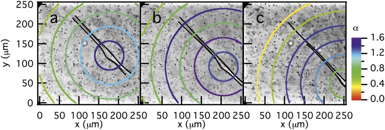
Fig. 4.
Dynamic spatial rheological data of the pericellular region during cell migration. Data are taken through time at (A) 0, (B) 24, and (C) 43 min after the cell is identified. This rapidly moving cell is causing the particles to move with the cell (outlined in black) as it migrates through the acquisition window. These measurements indicate that, once the cell is spread and begins to move, the scaffold is a viscoelastic fluid.

