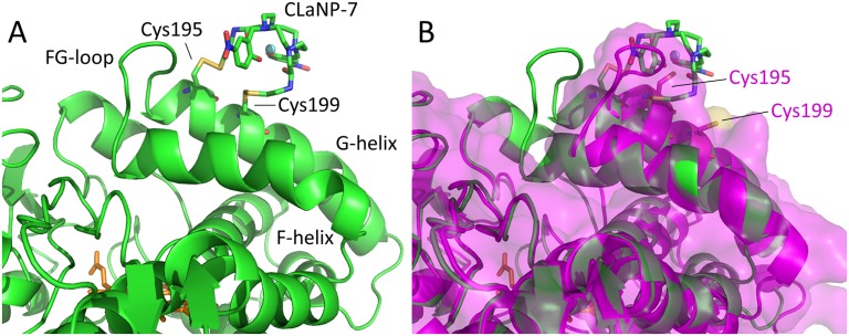
Fig. S3.
(A) Model of P450cam [camphor-bound, closed state; PDB ID code 3L63 (10)] with mutations 195/199C (cartoon), linked to CLaNP-7 (sticks). The Yb3+ ion is shown as a sphere. The heme is shown in orange sticks. CLaNP-7 was modeled to have the Yb3+ as close as possible to the origin of the experimentally determined tensor, with the z axis along the twofold symmetry axis of the CLaNP molecule. (B) Structure of A overlaid with the open state of camphor-free P450cam [PDB ID code 3L62 (10)] shown in magenta cartoons and a semitransparent surface, with mutations 195/199C modeled (sticks). CLaNP-7 shows severe steric clashes with the open state and is situated far from Cys199. It is concluded that the experimentally determined position of the probe shows that camphor-bound P450cam is in the closed state.

