Fig. 3.
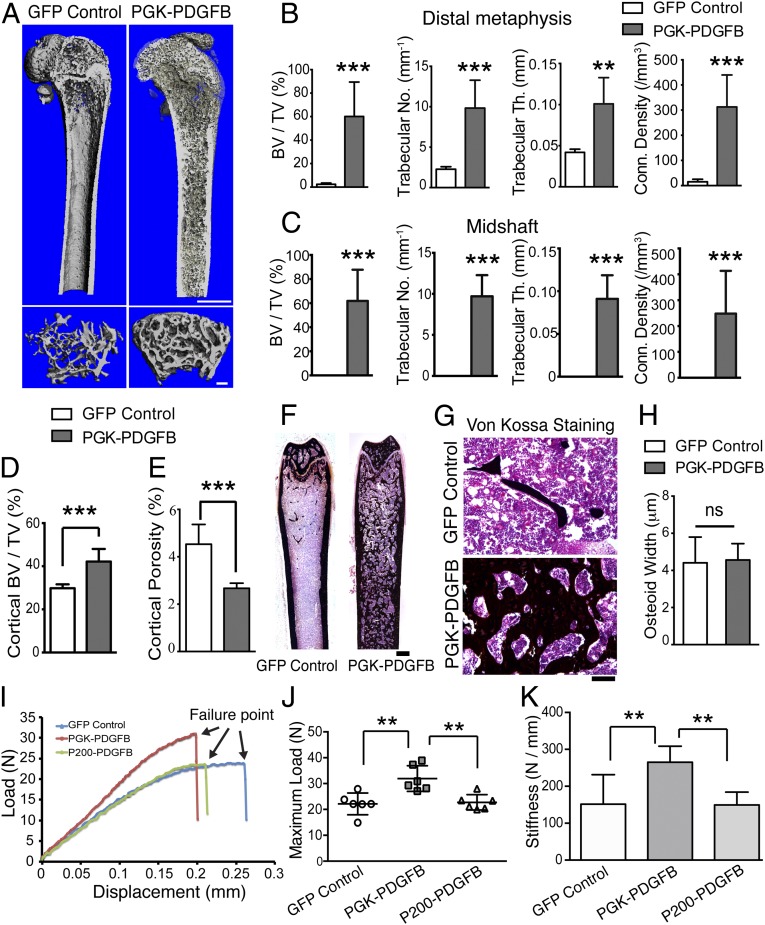
PGK-PDGFB treatment promotes de novo bone formation and enhances bone strength without causing osteomalacia. (A) The μCT 3D reconstruction images of a representative femur from the GFP control mice and the PGK-PDGFB–treated mice at 12 wk posttransplantation. (Upper) The bisected longitudinal image of the femur that shows trabecular structure within the marrow cavity. (Scale bar, 1 mm) (Lower) The cross-sectional area at secondary spongiosa of distal metaphysis of the femur. (Scale bar, 200 μm.) (B and C) Summary of the trabecular bone parameters at the distal metaphysis (B) and midshaft (C) of femurs of the PGK-PDGFB group and the GFP control group. n = 6 for each group. *P < 0.05. n = 6 per group. (D and E) Cortical bone volume (D) and cortical porosity (E) were determined at the midshaft of the PGK-PDGFB group and GFP control group. ***P < 0.001. (F and G) Von Kossa staining of femurs. Increased de novo bone formation in the marrow space in the Lenti-PGK-PDGFB group was observed. (Scale bars, 500 μm in F and 100 μm in G.) (H) Quantification of osteoid width (O.Wi). ns, not significant. (I) A representative loading force displacement curve of each test group. (J and K) Maximum load-to-failure (J) and stiffness (K) was significantly increased in the PGK-PDGFB–treated femurs compared with control or P200-PDGFB–treated bones. n = 6 per group. **P < 0.01.

