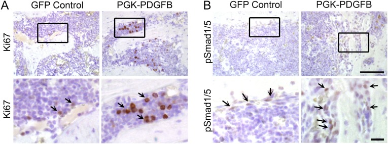
Fig. S9.
Ki-67 (A) and pSmad1/5 (B) immunohistochemical staining of the femur sections from control or PGK-PDGFB treated mice. The brown stain indicates the Ki-67 (arrows in A) and pSmad1/5 (arrows in B) proteins. [Scale bars, 100 μm in the low-magnification images (Upper) and 20 μm in the high-magnification images (Lower).]

