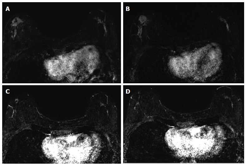
Figure 4.

Sixty-four years old woman with bilateral HR+ breast cancer. A and B: Baseline axial T1-weighted post-gadolinium fat-saturated magnetic resonance image demonstrates 3.2 cm irregular mass and contiguous non-mass enhancement (NME), spanning up to 7.2 cm, in the right central outer breast and 3.5 cm of clumped linear NME in the central outer left breast; C and D: Post-neoadjuvant chemotherapy magnetic resonance imaging demonstrates decrease in size of the right breast mass and NME. NME in the left breast demonstrates only mild improvement. Surgical pathology demonstrates 4.3 cm of residual disease on the right and 3.7 cm of disease on the left.
