
Fig. 4.
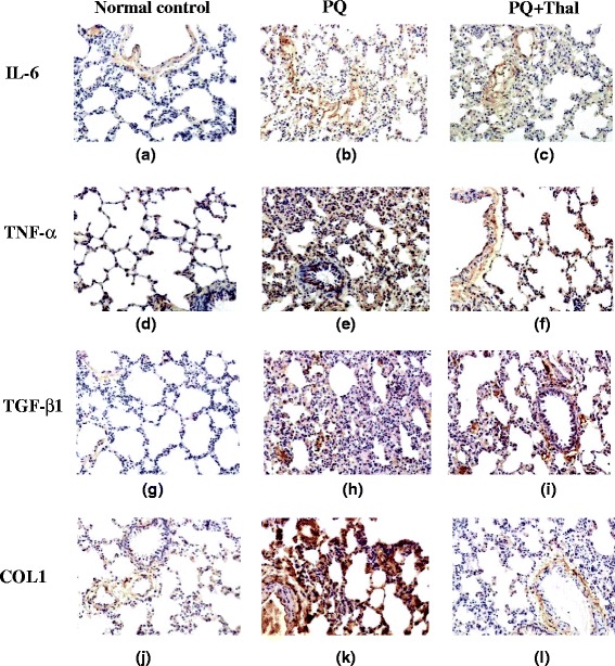
Immunohistochemistry detection of the expression of IL-6, TNF-α, TGF-β1 and COL1A1 proteins in lung tissues after PQ intoxication. Lung tissues from Control (a, d, g, j), PQ (b, e, h, k) and PQ + Thal (c, f, i, l) groups were collected on day 15 after PQ administration and expression of IL-6 (a, b, c), TNF-α (d, e, f), TGF-β1 (g, h, i) and COL1A1 (j, k, l) in lung tissues were examined using immunohistochemical staining (×100)