
Fig. 1.
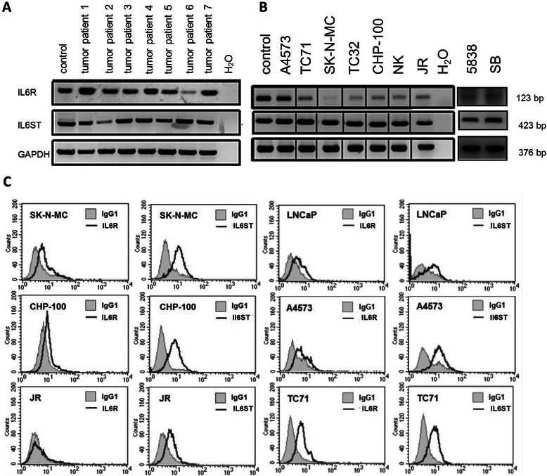
a IL6R and IL6ST mRNA expression of seven ES tumor samples by RT-PCR. IFN-γ treated PBMC were used as positive control (left lane). All tumor specimens expressed both IL6 receptor subunits. b IL6R and IL6ST mRNA expression in nine ES cell lines (RT-PCR). IFN-γ treated PBMC were used as positive controls (left lane). c Assessment of IL6 receptor complex cell surface expression by flow cytometry. The prostate cancer cell line LNCaP was used as a positive control. IL6R and IL6ST cell surface staining of 4 additional ES cell lines is shown in Additional file 1: Figure S1