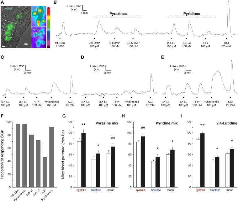
Figure 3.
Pyridine analogs are sufficient to mimic the systemic effects of predator urine in mice. (A) Coronal slice from the Grueneberg ganglion (GG) of an OMP-GFP mouse loaded with Fura-2AM. GG neurons (GGn) are localized between the nasal cavity (NC) and the nasal septum (NS). According to their intrinsic GFP fluorescence, GGn can be selected and observed either under Hoffman modulation view (Hv) or 380 nm in color-encoded map for unbound Fura. Here, a typical intracellular calcium increase induced by a control pulse of KCl (25 mM) observed before and at the peak in selected GGn (dashed rectangle). Scale bar, 10 μm in (A). (B) Representative continuous recording of a GGn responding to diluted urine of the mountain lion (Mt. Lion; 1:1000), to a set of pyrazines (2,6-dimethylpyrazine, 2,6-DMP; 2-ethyl-3,5-dimethylpyrazine, 2-EDMP; 2,3,5-trimethylpyrazine, 2,3,5-TMP; 100 μM) and to a set of pyridines (2,4-lutidine, 2,4-Lu; 3,4-lutidine, 3,4-Lu; 4-picoline, 4-Pi; 100 μM). (C–E) Examples of GGn with differential pattern of pyridine-evoked responses. (F) Proportion of responding GGn to the different tested cues. The Pyrazine mix (2,6-DMP, 2-EDMP, 2,3,5-TMP; 100 μM) and the Pyridine mix (2,4-Lu, 3,4-Lu, 4-Pi; 100 μM) were both able to initiate similar numbers of GGn responses comparable to the one observed with the diluted urine of the mountain lion (1:1000). A total of 60 viable GGn isolated from 6 mice (P1-7) were used (A–F). Fluorescence intensity Fura-2 ratio = F340/F380 is indicated by arbitrary units (a.u.); times are indicated by horizontal bars in (B–E). (G–I) Mice blood pressure analyzes by tail-cuff measurements for the Pyrazine mix (G), the Pyridine mix (H) or the 2,4-lutidine (I) at a dilution of 1%. Control conditions (white bars) and test sessions (black bars) are shown (G–I). Six adult male C57BL/6 mice were used (G–I). Values are expressed as mean ± SEM; one-tailed paired t-test or w-test, *P < 0.05; **P < 0.01; ns, not significant.

